
Percutaneous Intervention of Left Main Coronary Artery Chronic Total Occlusion
Abstract
Chronic total occlusion (CTO) of the left main coronary artery (LMCA) is rare on the angiograms; Coronary Artery Bypass Grafting is the standard method of its revascularization.
To demonstrate that PCI may in some cases be a safe option for patients with a high-risk surgical category, we report a complex clinical case of revascularization of chronic total occlusion of the LMCA, left anterior descending artery (LAD), and circumflex artery (CX).
Methods
Recanalization of the occluded LMCA and LAD was performed by utilizing the support-balloon technique, and CTO wires (Miracle 3™ wire, Abbott Vascular; Runthrough® NS Intermediate wire, Terumo); LAD, CX, LMCA, and its bifurcation, were stented with 3 drug-eluting stents (Resolute Integrity DES, Medtronic); the "Culotte Stenting " technique was used for bifurcation stenting, followed by "Kissing Balloon" post-dilatation technique; proximal optimization technique was performed in the LMCA.
Results
The intervention ended without complications. 2 months after stenting, the ejection fraction increased from 20% to 38%, improved almost all parameters of the heart, Congestive Heart Failure functional class decreased to class I.
Conclusions
It should be considered that LMCA CTO lesions can be successfully revascularized with PCI in case of the selection of the suitable patient and appropriate revascularization technique.
Author Contributions
Academic Editor: Elbaih Elbaih, Suez Canal University, Ismailia, Egypt.
Checked for plagiarism: Yes
Review by: Single-blind
Copyright © 2019 Murman Kantaria, et al.
 This is an open-access article distributed under the terms of the Creative Commons Attribution License, which permits unrestricted use, distribution, and reproduction in any medium, provided the original author and source are credited.
Competing interests
The authors have declared that no competing interests exist.
Citation:
Introduction
According to the World Health Organization, in 2015, 17.7 million people died from cardiovascular diseases (CVDs), with 75% of deaths occurring in developing countries and low-income countries. Nowadays 17.9 million people die each year from CVDs, an estimated 31% of all deaths worldwide 12. Cardiovascular diseases including coronary, cerebrovascular, peripheral arteries, as well as congenital and rheumatic heart diseases, deep vein thrombosis, pulmonary thromboembolism, are the leading cause of death worldwide.
It has been estimated that coronary chronic total occlusion (CTO) is encountered in 15 to 20% of patients referred for coronary angiography (CAG). Chronic total occlusion of left main coronary artery (LMCA) is rare on the angiograms and is described as a total lack of antegrade blood flow to the coronary arteries with retrograde collateral circulation 1, 11. Under these conditions, most of the myocardium is under ischemic stress, which is associated with high mortality. Although coronary artery bypass grafting (CABG) operation is the standard revascularization method, some recent studies testify that alternative percutaneous coronary intervention (PCI) can be successfully performed in a specific group of patients, and sometimes PCI is the only option for revascularization, especially for high-risk patients. The success of the PCI of CTO can be attributed to the vast array of devices that have now become available and to the vastly enhanced operator expertise. However, at the same time, it is necessary to note, that despite the tremendous success PCI, are recorded many cases of CTO, where PCI attempts failed 6
To demonstrate that PCI may in some cases be a safe option for patients with a high-risk surgical category, we report a complex clinical case of revascularization of chronic total occlusion of the LMCA, left anterior descending (LAD) artery, and circumflex (CX) artery.
Methods
Patient
The study was performed on the 62-year-old male patient with severe coronary artery disease (including chronic total occlusion of the left main coronary artery (LMCA)), previous myocardial infarction followed by ischemic cardiomyopathy, congestive heart failure of functional class III-IV (NYHA), periodically developing dyspnea and retrosternal discomfort (at rest or with minimal exertion), hydrothorax, peripheral edema, frequent hospitalizations.
CAG performed in 2014 revealed CTO of the LAD, moderately developed right-to-left collaterals, and an absence of significant stenosis in the right coronary artery (RCA).
The patient was treated at various clinics in Tbilisi (Georgia), was assessed as a high-risk patient, denied both to perform the intervention (stenting) as well as CABG, also had been denied implanting the Implantable Cardioverter-Defibrillator (ICD). Only medication treatment was recommended. The patient's prognosis was - a candidate for sudden cardiac death.
In May 2017, a patient was hospitalized at the “Aversi” Clinic due to acute heart failure. The patient underwent the Ultrasonography examination and the SYNTAX score was calculated.
Recanalization
Recanalization of the occluded LMCA and LAD was performed by utilizing the support-balloon technique, and CTO wires (Miracle 3™ wire, Abbott Vascular; Runthrough® NS Intermediate wire, Terumo); LAD, CX, LMCA, and its bifurcation, were stented with 3 drug-eluting stents (Resolute Integrity DES, Medtronic); the "Culotte Stenting" technique was used for bifurcation stenting, followed by "Kissing Balloon" post-dilatation technique; proximal optimization technique was performed in the LMCA.
Results and Discussion
Ultrasound examination of the patient revealed cardiac ejection fraction (EF) - 20% (versus 55% or more in control), diffuse hypokinesis, dilated left ventricular diameters (end-diastolic diameter, 60 mm; end-systolic diameter, 51 mm), severe (restrictive) diastolic dysfunction, mild mitral and tricuspid regurgitation, trivial aortic regurgitation.
The SYNTAX score was calculated to be 47.
After medical treatment and comparable improvement in the patient's condition, expected risks were re-evaluated. After consulting with the patient and family members (they did not agree to the operation based on past experience) it was resolved to perform repeated CAG and to attempt revascularization of the LAD artery, (stenting). However, repeated CAG additionally revealed chronic 100% occlusion of the LMCA (Figure 1), and also CX occlusion, which was unexpected, and thereby significantly decreasing the likelihood of procedural success. In this case, the patient's heart was supplied only through the right coronary artery, which partially supplies the occluded left anterior descending and circumflex arteries through the contralateral collateral blood flow (Figure 2). Acute LMCA occlusion in most of the cases is fatal and survival is possible only in patients with a dominant RCA providing sufficient collateral formation.
Figure 1.Chronic total occlusion of the left main coronary artery
Figure 2.Right Coronary Artery with developed right-to-left collaterals.
In spite of the unexpected difficulties, after consultations with the patient's family members, the intervention was resolved because, without improvement of the coronary blood supply, the patient was doomed to sudden cardiac death soon.
The goal of the intervention was partial or complete revascularization, improving the contractile function of the heart.
The plan of the intervention included:
1. Revascularization of chronic total occlusions of the LMCA and CX;
2. Revascularization of CTO of the LAD artery;
3. Stenting of the LAD artery, circumflex artery, left main coronary artery, and its bifurcation.
All three of the steps listed above separately represent the challenging, time-consuming and high-risk procedures. Considering the patient's condition (heart's ejection fraction of 20%, which means that only 1/3 of the coronary blood supply is preserved), simultaneous implementation of all three stages presents significant technical difficulty and poses a high risk for the patient, as any significant complications or procedural misconduct will result in cardiac arrest and death.
The procedure was continued for 2 h 30 min. To prevent pulmonary congestion and heart failure, the patient was in the "Reverse Trendelenburg " position on the operating table, a forced diuresis was performed, and a resuscitation team was mobilized. PCI was performed with only radial artery access, using a 6fr sheath, and 6fr Extra Backup coronary guide catheter (Launcher, Medtronic).
In the first stage, occluded left main and circumflex arteries were recanalized utilizing the support-balloon technique, and CTO wires (Miracle 3™ wire, Abbott Vascular; Runthrough® NS Intermediate wire, Terumo) and balloon pre dilatation was done (Figure 3). In the second stage, the occluded LAD artery was successfully recanalized by utilizing the support-balloon technique, and CTO wires (Figure 3) (due to severe calcinosis the second stage lasted 40 min). In the third stage, LAD artery, CX artery, as well as LMCA and its bifurcation, were stented with 3 drug-eluting stents (Resolute Integrity Zotarolimus DES, Medtronic), the "Culotte Stenting " technique was used for bifurcation stenting (Figure 4), followed by "Kissing Balloon" post-dilatation technique, and finally, proximal optimization technique was performed in the LMCA. The final angiographic image is good, the intervention ended without complications (Figure 5).
Figure 3.LMCA, CX, LAD after recanalization and predilatation
Figure 4.Culotte stenting technique
Figure 5.Left Coronary Artery, final result of the intervention.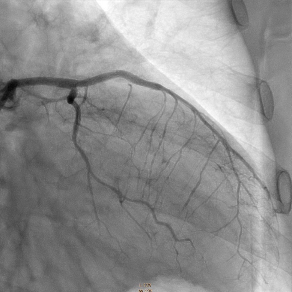
2 months after PCI, the ejection fraction increased from 20% to 38%, improved almost all parameters of the heart 8, 10 (Fractional Shortening (FS) increased from 15% to 25%; Left ventricular end-systolic dimension (LVESD) decreased from 51 mm to 38mm; left ventricular end-diastolic diameter (LVEDD) decreased from 148 mm to 89 mm; left atrium (LA) size decreased from 44 mm to 41mm; Right Ventricle (RV) size decreased from 38 mm to 35mm; Right Atrium (RA) size decreased from 49 mm to 38mm; Systolic Pulmonary Artery Pressure (PAPs) decreased from 64 mm/Hg to 21mm/Hg;) (see Table 1); the functional class of the Congestive Heart Failure decreased from class III-IV (Moderate-Severe) to class I (Mild), and the patient no longer required ICD implantation.
Table 1. Patient’s heart parameters before and after the intervention| Parameter | Before Intervention | After Intervention |
| EF | 20% | 38% |
| Fractional Shortening (FS) | 15% | 25% |
| LVESD( Left Ventricular End-Systolic Dimension) | 51 mm | 38 mm |
| LVEDD(left ventricular end diastolic diameter) | 60 mm | 52 mm |
| LVEDV( left ventricular end-diastolic volume) | 148 mm | 89 mm |
| left atrium (LA) | 44 mm | 41 mm |
| Right Ventricule (RV) | 38 mm | 35 mm |
| Right Atrium (RA) | 49 mm | 38 mm |
| Systolic Pulmonary Artery Pressure (PAPs) | 64mm/Hg | 21 mm/Hg |
Revascularization of LMCA CTO in patients with the use of PCI and CABG methods is associated with a certain procedural risk. Long-term results of CABG operation in LMCA CTO patients with a high SYNTAX score usually give a better prognosis, but, as shown, there is no significant difference in the long-term results of using these two methods (PCI and CABG), if the selected procedure is suitable for the patient 5, 7. A few, but promising results were obtained after the use of new-generation drug-eluting stents 2, 9. New tools and techniques have allowed interventional cardiologists to adjust more and more complex lesions, but for the treatment of LMCA CTO, PCI is very rarely used.
In the case presented above, although the high SYNTAX score would have indicated the need for surgical revascularization (CABG), because of the poor functional status (NYHA III-IV class) we found, that PCI has low-risk options compared to surgery and in case of success could provide the optimal remote result.
In the literature available to us have been reported only a few cases using the Intravascular ultrasound (IVUS) guidance - bilateral femoral assess, and large diameters guide catheters 3, 4. Our case is unique because we have achieved an optimal result without IVUS guidance, using only single radial artery asses, and comparably small-sized single guide catheter (6Fr). The final angiographic result was good, however, we could not confirm the optimal result with IVUS due to several technical and financial (nor covered by insurance) problems in our hospital.
Conclusion
As a result of interventional measures and optimal medication treatment
The patient received a complete revascularization.
The quality of life has improved significantly
The risks were significantly reduced and the forecast improved
More than two years after the intervention, the patient has no complaints (according to MACE), there was no need for hospitalization, the patient performs regular physical activity without angina, echocardiographic data remained unchanged, ejection fraction ranges from 38-40%.
It should be concluded that LMCA CTO lesions may be successfully revascularized with PCI by appropriate patient and revascularization technique selection, we believe that IVUS guidance is not obligatory, but it is very important for proper stent sizing, deployment, and also it is important to confirm final optimal result.
Abbreviation: CABG - coronary artery bypass grafting; CAG - coronary angiography; CTO - chronic total occlusion; CVDs cardiovascular diseases; PCI - percutaneous coronary intervention; LMCA - left main coronary artery; LAD - left anterior descending artery; CX - circumflex artery; RCA - right coronary artery; ICD - Implantable Cardioverter-Defibrillator; IVUS - Intravascular ultrasound; NYHA - New York Heart Association; EF – Ejection Fraction; FS - Fractional Shortening; LVEDD - left ventricular end-diastolic diameter; LVESD - Left Ventricular End-Systolic Dimension; PAPs - Systolic Pulmonary Artery Pressure;
References
- 1.Akhtar R P, Naqshband M S, Abid A R, Tufail Z, Waheed A et al. (2009) Surgery for chronic total occlusion of the left main stem: A 10-year experience. Asian Cardiovasc Thorac Ann. 17, 472-476.
- 2.Bardají A, Rodriguez-López J, M. (2014) Torres-Sánchez. Chronic total occlusion: to treat or not to treat. , World J Cardiol 6, 621-629.
- 3. (2018) Çoksevim Metin, Murat Akçay, Korhan Soylu, Ömer Göktekin. Successful Percutaneous Intervention of Left Main Coronary Artery Chronic Total Occlusion:A Case Report. , Anatol J Cardiol 19(6), 419-421.
- 4.Banning. (2013) Intravascular ultrasound-guided revascularization of a chronically occluded left main coronary artery. , Indian Heart J 65.
- 5.S De Rosa, Polimeni A, Sabatino J, Indolfi C. (2017) Long-term outcomes of coronary artery bypass grafting versus stent-PCI for unprotected left main disease: a meta-analysis. , BMC Cardiovasc Disord 17, 240.
- 6.Dash Debabrata. (2018) Interventional Management of “Balloon-Uncrossable” Coronary Chronic Total Occlusion: Is There Any Way Out? Korean Circ J. 48(4), 277-286.
- 7.Giacoppo D, Colleran R, Cassese S, Frangieh A H, Wiebe J et al. (2017) Percutaneous Coronary Intervention vs Coronary Artery Bypass Grafting in Patients With Left Main Coronary Artery Stenosis: A Systematic Review and Meta-analysis. , JAMA Cardiol 2, 1079-1088.
- 8.Lancellotti P, Badano L P, Lang R M, Akhaladze N, Athanassopoulos G et al. (2013) Normal reference ranges for echocardiography: Rationale, study design, and methodology (NORRE study). Eur Heart J Cardiovasc Imaging. 14(4), 303-308.
- 9.G Z Ma, Wang Y, R H Xu. (2016) Successful percutaneous coronary intervention for acute and chronic occlusion of the left main coronary artery: report of two cases and review of the literature. , Acta Cardiol 71, 127-134.
- 10.Morice M C, Serruys P W, Kappetein A P, Feldman T E, Ståhle E et al. (2010) Outcomes in patients with de novo left main disease treated with either percutaneous coronary intervention using paclitaxel-eluting stents or coronary artery bypass graft treatment in the Synergy between Percutaneous Coronary Intervention with TAXUS and Cardiac Surgery (SYNTAX) trial. , Circulation 121, 2645-2653.