
Fetal Abdominal Cystic Lesion: A Diagnostic Dilemma and Prognostic Challenge-Report of Two Cases of Mesentric Lymphangioma with Review of Literature
Abstract
Fetal mesentric lymphangioma, a congenital fetal abdominal cystic malformation has a rare occurrence. Antenatal detection, its differential diagnosis, prenatal management options and parental counseling regarding postnatal prognosis of such a case are thus rarely discussed. We report two cases of antenatally detected fetal cystic abdominal mass with a provisional diagnosis of abdominal lymphangioma. Postnatally one of the neonates developed features of intestinal obstruction and required surgical intervention. Intra-operative findings and histopathology report confirmed a mesentric lymphangioma. The other neonate had associated subcutaneous lymphangiectasia in left lumbar region, left sided inguinal hernia, undescended testes and was asymptomatic postnatally and managed conservatively.
Author Contributions
Academic Editor: HASSAN ABDULJABBAR, King Abdulaziz University, Jeddah, Kingdom of Saudi Arabia
Checked for plagiarism: Yes
Review by: Single-blind
Copyright © 2021 Daniel Aboma, et al.
 This is an open-access article distributed under the terms of the Creative Commons Attribution License, which permits unrestricted use, distribution, and reproduction in any medium, provided the original author and source are credited.
Competing interests
The authors have declared that no competing interests exist.
Citation:
Introduction
The detection rate of fetal abdominal cysts and other anomalies is increasing with wide spread use of antenatal ultrasonography. These cysts usually arise from gastrointestinal or genitourinary system and are more frequent and larger in size in a female fetus as compared to male fetus1. Fetal abdominal lymphangioma is reported by very few authors. Site, extension, pressure symptoms, other malformations or syndromic associations determine prognosis of a lymphangioma2. Fetal abdominal cystic lesions can be detected from late first trimester ultrasonography scan but, it is difficult to make a precise diagnosis antenatally. A comprehensive analysis by an expert radiologist regarding the site of origin, nature of the cyst and follow-up scans might guide to the diagnosis. Magnetic resonance imaging in doubtful cases could help in prognosticating and counseling the parents regarding the morbidity and mortality rates3. We report two cases of antenatally detected mesenteric lymphangiomas, in which one of the fetuses presented with intestinal obstruction and operated in the neonatal period and other was asymptomatic till one year of age and was managed conservatively. Diagnostic dilemma and prognostic challenge of various fetal abdominal cystic lesions in pregnancy has been discussed in detail along with review of literature in this report.
Case Report
Case 1
24years old primigravida reported to obstetric emergency room at 37weeks period of gestation in labor. Her antenatal period was supervised at private hospital and was apparently uncomplicated, an obstetric scan done elsewhere in her third trimester reported a single live fetus with a fetal abdominal cystic mass measuring 6.3 x 5 cm and rest of the fetal organs were normal with normal growth parameters. Antenatal counseling and prognosis regarding the nature of lesion was not provided to her from her visiting hospital. Exact diagnosis was not feasible in the intrapartum period; nevertheless a provisional diagnosis of fetal abdominal mesenteric cyst was made. Labor progressed well and she had normal delivery of a live born male weighing 1.8 kilograms, there were no obvious gross malformations in the newborn and apgar scores were normal. Neonate was apparently healthy and was well accepting breastfeeds till day two of life following which he developed features of intestinal obstruction. He was taken up for emergency laparotomy and intraoperatively there was a lobulated cystic mass around 4 x 5 cm in diameter in relation to the jejunum (Figure 1) as the cause for obstruction, which was excised along with jejunal resection (4cm of jejunum) and jejunojejunal anastomosis. Postoperative period has been uneventful and baby was discharged in healthy condition. The histopathology report confirmed the cyst as lymphangioma of bowel mesentery.
Figure 1.Fetal Abdominal Mesentric Lymphangioma along with part of jejunum.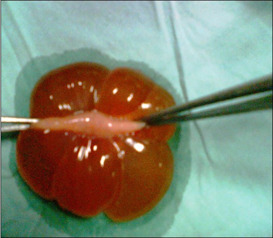
Case 2
34 year old G2P1001 was referred to our institute at 21weeks period of gestation for a fetal abdominal cystic mass for further evaluation and management. At referral, she had an anomaly scan reporting a left sided retroperitoneal collection of 3x2 centimeters with septations surrounding the kidney and extending up to left pelvis suggestive of lymphocoele in the fetus. In second trimester anomaly scan nuchal fold thickness was normal, there were no markers suggestive of aneuploidy and no other malformation was seen. In follow-up scans, there was increase in size of the collection upto 8x6 cm at term which also shown a subcutaneous component of around 5x4 cm along the left fetal torso in the lumbar region (Figure 2a). Growth parameters were normal and amniotic fluid was adequate. She was hypothyroid on treatment and developed gestational diabetes at 32weeks period of gestation which was managed with medical nutrition therapy. Her first child was born by cesarean section and had no congenital malformation. Index pregnancy was terminated by an elective cesarean section at term as she was not willing for vaginal birth after caesarean. Male baby of 3.4kg was born with good apgar score. He had a left sided lumbar cystic swelling of 5x6cm, reducible inguinal hernia and undescended testes on the left side. Postnatal echocardiography was normal and postnatal USG shown similar features of the cyst. Baby was active, euthermic, euglycemic, passed urine and stool and was well accepting feeds hence discharged at day 3 of life. He was conservatively followed and at 6 months postnatal visit, size of the lumbar swelling was clinically decreased in size and infant had age appropriate milestones. Ultrasonography (Figure 2b) and CECT scan at one year of age was suggestive of large cystic lesion in the abdomen and pelvis (13*6*12 cm) with extension into postero lateral subcutaneous planes of abdominal wall through a defect in the left oblique muscle. Rest of the abdominal organs and large vessels were normal and there was no ascites or lymphadenopathy. However child is doing well at one year of age without any clinical obvious increase in the size of lumbar swelling.
Figure 2a.Images of Ultrasonography at second trimester of pregnancy
Figure 2b.Images of Ultrasonography at one year of age.
Discussion
Fetal abdominal cystic lesions occur in isolation in around two-third of cases, and in other one-third there may be associated anomalies. Presence of associated anomalies, oligohydramnios or polyhydramnios, increased nuchal translucency in a first trimester scan, dysmorphic facies increase the probability of fetal aneuploidy.4, Table 1. demonstrate causes for various fetal abdominal cysts.5 Diagnostic accuracy may be increased with knowledge of fetal gender, site and correlation with adjacent organs. Serial monitoring is essential as most of the cysts regress spontaneously.6
Table 1. Causes of fetal abdominal cysts5.| 1.Urogenital cystic lesions (40%) | Hydronephrosis | Secondary to Ureteropelvic Junction Obstruction may be associated with central nervous system , cardiovascular system , gastro-intestinal, skeletal malformations and aneuploidies, trisomy 21 commonly. |
| Bladder outflow obstruction secondary to posterior urethral valves-sporadic or familial disorder. Associated abnormalities may be tracheo-esophageal fistula, total anomalous pulmonary drainage, mitral stenosis, skeletal abnormalities, imperforate anus and trisomy 13,18. | ||
| Renal dysplasia | Multicystic dysplastic kidney may occur in association with many syndromes like Dandy-Walker and Apert's syndromeZellweger's, Meckel-Gruber and Jeune syndrome are associated with nonobstructive cystic dysplasia | |
| Urachal cyst | Can be as an isolated anomaly or in association with prune-belly syndrome. | |
| Ovarian cysts | Polyhydramnios is seen in 5-10%of patients. Complications include torsion, hemorrhage, necrosis, rupture and intestinal obstruction. | |
| Hydrometrocolpos | Secondary to imperforate hymen or as a part of McKusick- Kaufman syndrome | |
| 2.Gastrointestinal cystic lesions (15%) | Duodenal atresia or stenosis | May have other intestinal atresias, congenital heart disease and the VACTERL association. About 30% of cases are associated with Down's syndrome. |
| Small bowel obstruction | Usually associated with polyhydramnios.Abnormal karyotype is rare. | |
| Large bowel obstruction | Hirsch-prung disease- Down's syndrome is seen in around 5% with these. | |
| Duplication cysts | May be associated with developmental disorders of spinal cord and blood vessels. | |
| Mesenteric and Omental cysts | lymphatic hamartomas, described in detail in discussion. | |
| 3. Other Cystic mass. | Precise antenatal diagnosis of Cystic mass arising from hepato-biliary system, adrenal glands, pancreas, spleen other abdominal organs and retroperitoneum is challenging. |
Fetal abdominal cystic masses usually arise from ovary or gastrointestinal tract. Cysts from gastrointestinal tract (GIT) may be omental or mesenteric in origin and or may arise consequent to intestinal obstruction, gastric duplication. The usual presentation of mesentric cyst is a single, multilocular cyst which may vary in size from less than a centimeter to a huge one. Symptoms vary depending upon the size of the cyst and the content within the cyst may be serous, chylous or hemorrhagic in nature. These should be differentiated from cysts arising from ovary and retroperitoneum.Massive cysts or those appear grave with other major congenital malformations and/or aneuploid fetuses, medical termination of pregnancy could be a reasonable approach, provided gestational age at diagnosis meets the period of legality for medical termination of pregnancy of the country. Surveillance for single, small, innocent cysts can be done by serial ultrasonography and the pattern of progression or regression can be noted. Management either in the prenatal or postnatal period can thus be planned accordingly. Case 1 presented as isolated non lethal cyst antepartum nevertheless need to be operated for intestinal obstruction in immediate neonatal period. Case 2 presented as multiseptated slowly growing fetal abdominal cyst with subcutaneous extension and did not require any surgical intervention till one year of followup.
Lymphangioma is a hamartomatous malformation or developmental defect arising due to interruption in communication of embryonic lymph sacs with the venous channels. They have an incidence of 1 in 6000 live-births. These appear as unilocular or multilocular benign cystic mass in head and neck region with a potential to infiltrate into the surrounding viscera. Lymphangioma can be of three types: lymphangioma simplex, cavernous type, and cystic hygroma or lymphangioma. They can originate from any of the lymphatic channels; around 95% occur in the neck (usual site of representation for Cystic hygroma), head or axilla. Cystic hygroma is usually associated with Turners syndrome with XO fetal karyotype. Abdominal lymphangiomas usually arise from small bowel mesentry or from retroperitoneum and can extend upto lower limbs7,8. The size may be variable as a small encysted fluid collection to a complex, huge cyst9. USG is the diagnostic modality of choice. The classical appearance of Lymphangioma on ultrasonography is a hypoechoic mass with thin walls and multiple septations. However, antenatal magnetic resonance imaging (MRI) scans provide a detailed view of anatomical relationship of the cysts to the surrounding tissue complementing characteristic USG images10.
Spontaneous regression is rare and mesenteric lymphangiomas depending on its location and locoregional spread usually require surgical intervention after birth.7,8 Conservative approach like cystic drainage is not recommended because of their tendency to recur irrespective of the modality chosen. Prognosis is good after surgical excision but follow-up is advised for possibility of recurrence. The postoperative morbidity for diffuse and multiple lesions is high as the procedure involves partial bowel obstruction and anastomoses. Case 1 also had resection of part of the jejunum along with removal of the cyst, and there is no recurrence at five years of follow up. Adjuvant therapy withbiological response modifier with antineoplastic effects like OK-432, are emerging as a potential therapy in preventing further enlargement of small localizedcysts9.
Thus favourable prognostic factors for lymphangiomas are : a single isolated lesion with no extension to lower limbs or surrounding structures, quiescent or slow growth, first appeared in third trimester, not situated in posterior neck and normal karyotype. A multidisciplinary approach with thorough antenatal evaluation and parental counseling with neonatologists and pediatric surgeons is recommended to achieve optimum perinatal outcome.
References
- 1.Husen M, Schut P, A C Neven, C H, Yousoufi N et al. (2019) . Differences in Origin and Outcome of Intra-Abdominal Cysts in Male and Female Fetuses. Fetal Diagn Ther 46, 166-174.
- 2. (2013) The value of early recognition of fetal lymphangioma. Farnaghi and Kothari. , AJUM August 16(3).
- 3.Tu C Y. (2017) Ultrasound and differential diagnosis of fetal abdominal cysts. Exp Ther Med. Jan;13(1): 302-306. doi: 10.3892/etm.2016.3948. Epub 2016 Dec 2. PMID: 28123506; PMCID: PMC52451335 .
- 4.Stirnemann J MarchitelliG, Rousseau V AcanforaMM, Salomon L J, Ville Y. (2015) Prenatal diagnosis of intra-abdominal cystic lesions by fetal ultrasonography: diagnostic agreement between prenatal and postnatal diagnosis.PrenatDiagn. 35(9), 848-52.
- 5.Rajesh Agarwal. (1999) Sonographic assessment of fetal abdominal cystic lesions: A pictorial essay.Indian. , Journal of Radiology and Imaging 9, 169-82.
- 6.Sahiti Mattegunta, Keerthy Rethiman M, Andrew Chitra. (2019) Fetal Abdominal Cyst. , IOSR Journal of Dental and Medical Sciences (IOSR-JDMS) 18(1), 1-4.
- 7.Açıkgöz A S, Tüten A, Bulut B, Öncül M, Eskalen Ş. (2015) . Fetal Abdominal Cysts: Prenatal Diagnosis and Management. Gynecol Obstet (Sunnyvale) 5, 1-4.
- 8.OzyuncuO1 CanpolatFE, CiftciAO YurdakokM, OnderogluLS DerenO. (2010) Perinatal outcomes of fetal abdominal cysts and comparison of prenatal and postnatal diagnoses.Fetal DiagnTher. Epub 28(3), 153-9.